
Case presentation
It is a sequel from a talipes equinovarus operated initially by a classic Achilles tendon lengthening technique to correct the deformity of the foot. The partial atrophy of the leg concerns the bones, the joints and the muscles. It does not induce major functional discomfort but a slight limp and above all a social life complex: stare discomfort, clothing restrictions, team sports apprehensiveness.
The two legs asymmetry is clear from the front and the back. The patient strongly wishes a surgical correction.
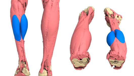
We offer him a volume increase of the two posterior calf muscles (gastrocnemius or twins) by an unalterable silicone custom-made implant. The implant is designed from a CT scan of both legs using computer-aided design (CAD) giving an improved anatomical image of both muscles but without obtaining a perfect symmetry due to the associated bone atrophies related to the pathology.

Surgery
The operation
48h-hospitalisation, 30min-operation :
- the patient is intubated in prone position under general anaesthesia,
- an around 3inches-horizontal-skin-incision in the popliteal fossa of the knee,
- opening of the aponeuroses of the twin muscles on each side,
- detachment of the avascular space between muscle and aponeurosis on the exact surface of the implant,
- introduction of the semi-rigid implant on each side respecting the vertical partition separating the two muscles,
- closure in two sections with absorbable suture with intradermal suture. No drain,
- moderate circular compression of the leg.
Operation follow-up
The patient gets up the next day with the help of a physiotherapist, walks with a forearm crutch and leave the hospital the same evening under analgesics (paracetamol). For 8 days, analgesics, gentle circular compression, walking, legs raised in sitting or lying positions. Follow-up consultation 8 days later and three months for resumption of sport.
Outcome
Efficient and rapid operation, moderate risks, objective improvement of the deformity without perfect symmetry (Fig. 3). The implants are placed for life with no risk of rupture, retractable shell or displacement. There can't be a "rejection".

Author and bibliography
Author
Former head of the plastic surgery department at Toulouse University Hospital, Prof. Chavoin has specialized for the last 15 years in the correction of malformations by custom-made 3D silicone implants. Concerning calf atrophy, he was inspired by the work of Glicenstein and adapted its work with personalised silicone implants.

