Mr Ian Hunt and Mr Ashok Kar, cardio-thoracic and Pectus experts
Mr Ian Hunt, BSc (Hons) MBBS MRCS FRCS (CTh), is the Medical Director of the Pectus Clinic, a Thoracic Surgeon and Clinical Lead Consultant for the Department of Thoracic Surgery, Division of Cardiothoracic and Vascular Surgery, at St. George’s Hospital NHS Trust and Spire St. Anthony’s Hospital.
He has an active interest in thoracic trauma and is an internationally recognized expert on chest wall and pectus deformities. He is actively involved in clinical research, training and education at undergraduate and postgraduate level. He has over 150 presentations, articles and publications and has talked at many national and international meetings on a wide variety of thoracic surgery topics. He has written reviews and book chapters and has edited several books.
Mr Hunt, working with Mr David Gateley, an experienced Plastic Surgeon and under the guidance and support of Prof Chavoin has performed nearly 50 pectus implants procedure, with around of quarter of patients (25%) having had a previous pectus operation (Nuss or modified Ravitch).
Mr Ashok Kar, BA (Hons), MB BChir, MA (Cantab), MRCS (Eng), FRCS (CTh), is a Cardio-Thoracic Surgery Specialist Training Registrar (ST8), and London Postgraduate School of Surgery and St George’s Hospital NHS Trust.
He is a senior cardio-thoracic surgery registrar (resident) with an interest in trauma and chest wall deformities. He has been awarded multiple national prizes during his training in cardio-thoracic surgery and has presented internationally at both the American Association for Thoracic Surgery and European Society for Thoracic Surgery Annual Meetings on innovative techniques related to the Nuss Procedure.
Clinical case : Implants after hammock technique failure for Pectus Excavatum
Clinical scenario
Sex: Male
Age: 22 years old
Context: Previous modified Ravitch surgical correction with hammock technique for severe asymmetric Pectus Excavatum
Presentation
This young male patient had no physical symptoms from his pectus excavatum but had noticeably found the cosmetic appearances more challenging as he reached adolescence.
He was initially investigated with lung function and echocardiogram, which did not demonstrate any significant physiological impairment. He had no other notable past medical history.
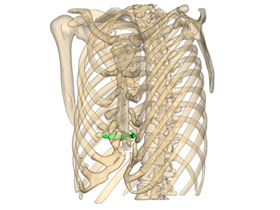
In 2017 he proceeded to undergo surgical correction of his severe pectus deformity with a modified Ravitch approach, using a prolene mesh sling technique rather than metal bar to maintain the sternal correction. The technique involves using a length of prolene mesh rolled into a tube and then placed under the freed-up sternum as its lowest point. The mesh is then securely anchored to the rib cage on either side having been pulled tight to lift the sternum. The sling or hammock technique was first described by Robicsek[1999, ref. 1].
After cartilage resection during a Ravitch-type-procedure the sternum is suspended with a hammock consisting of a Marlex mesh. The technique became popular amongst thoracic surgeons as it does not require a 2nd operation to remove a metal bar typically used to support the sternum after a modified Ravitch procedure.
Following the surgery, he made an uneventful recovery, but the patient reported a rapid recurrence of his pectus excavatum within in a few weeks. The patient was referred for a second opinion with residual moderate to severe, relatively asymmetric, and rightward pectus excavatum.
Investigations
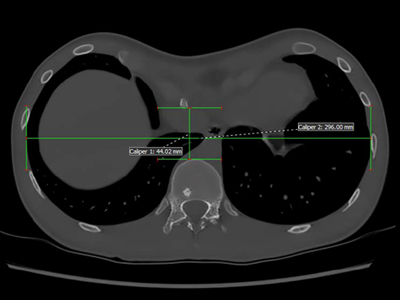
A repeat CT scan was performed which demonstrated a Haller index of 6.7, despite previous surgical correction of the pectus excavatum deformity.
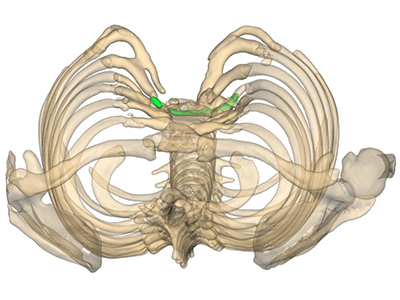
Detailed Chest reconstructions were completed as part of his assessment. The patient was keen to avoid further revision corrective surgery which would have required a revision modified Ravitch procedure and removal of the prolene mesh with increased risks of complications due to uncertain intrapleural scarring and adhesions behind the sternum.
Proposed Further Treatment
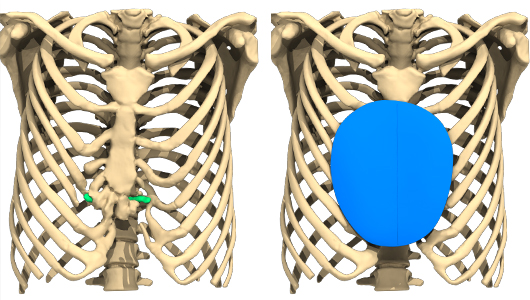
The patient opted for a custom-made 3D silicone implant with his surgeon. The implant was specifically designed for the patient according to the 3D bone and muscle reconstruction of CT images. A fairly small discrete (458cc by volume) but quite deep implant (4cm in depth) was rendered and manufactured.
Surgical Implant Procedure
The patient underwent their pectus implant operation in April 2018.
This was performed as a day case procedure with dedicated thoracic surgeon and plastic surgeon. The patient was measured, and the implant template used to mark the position of the template against bony landmarks and according to Anatomik Modeling instructions.
A transverse sternal incision was made through the previous incision over the deformity. The soft tissues including pectoralis major were dissected off the deformity bilaterally.
A pocket between the pectoralis muscle superficially and sternum and anterior chest wall deep was created. Haemastasis was secured and the implant placed with an excellent fit and secured to soft tissues. The patient had an uneventful post-operative recovery and was discharged the same day, with a Redivac Drain. This was subsequently removed 12 days later during clinic follow-up.
Results
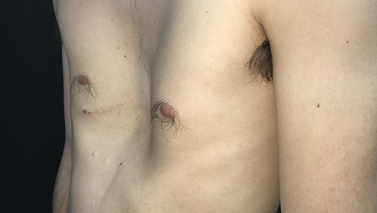
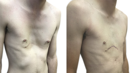
An excellent cosmetic result to correct the pectus deformity and chest wall asymmetry using the implant was achieved. The patient was entirely happy with the final result and discharged from further follow-up after 3 months.
Left Figure : Photos taken two months after pectus implant procedure to correct the residual pectus excavatum.
Patient Testimonial
“After having a Ravitch……I was still affected by my pectus and let it hold me back in life. Having had a pectus implant I feel normal and very glad I decided to go ahead”
Conclusions
Occasionally the result following a corrective operation can be unsatisfactory – a modified Ravitch technique that utilises a sling or hammock technique may avoid an 2nd operation to remove the metal bar but may be associated with a risk of early recurrent pectus excavatum deformity. Redo surgery in these situations can be associated with greater operative risks and post-operative complications.
This case highlights that a pectus implant can be used in this clinical setting with excellent outcomes, uneventful recovery and no need for further intervention.
Source and Bibliography
Source
Bibliography
Patel AJ, Hunt I. Initial Reduction of flexible Pectus Carinatum with Outpatient Manipulation as an adjunct to External Compressive Bracing: Technique and Early Outcomes at 12 weeks. J Pediatr Surg. 2019 Nov 1. pii: S0022-3468(19)30672-4. doi: 10.1016/j.jpedsurg.2019.09.024. [Epub ahead of print] PubMed PMID: 31708203.
Fraser S, Harling L, Patel A, Richards T, Hunt I. External Compressive Bracing with Initial Reduction of Pectus Carinatum: Compliance is the Key. Ann Thorac Surg. 2019 Sep 23. pii: S0003-4975(19)31411-0. doi: 10.1016/j.athoracsur.2019.08.026
Patel AJ, Hunt I. Is Vacuum Bell therapy effective in the correction of pectus excavatum? Interact Cardiovasc Thorac Surg. 2019 Mar 28. pii: ivz082. doi: 10.1093/icvts/ivz082.
Patel AJ, Hunt I. Effectiveness of Compressive External Bracing in Patients with flexible Pectus Carinatum Deformity: A review. Thorac Cardiovasc Surg. 2019 Apr 25. doi: 10.1055/s-0039-1687824. [Epub ahead of print] PMID: 31022736

